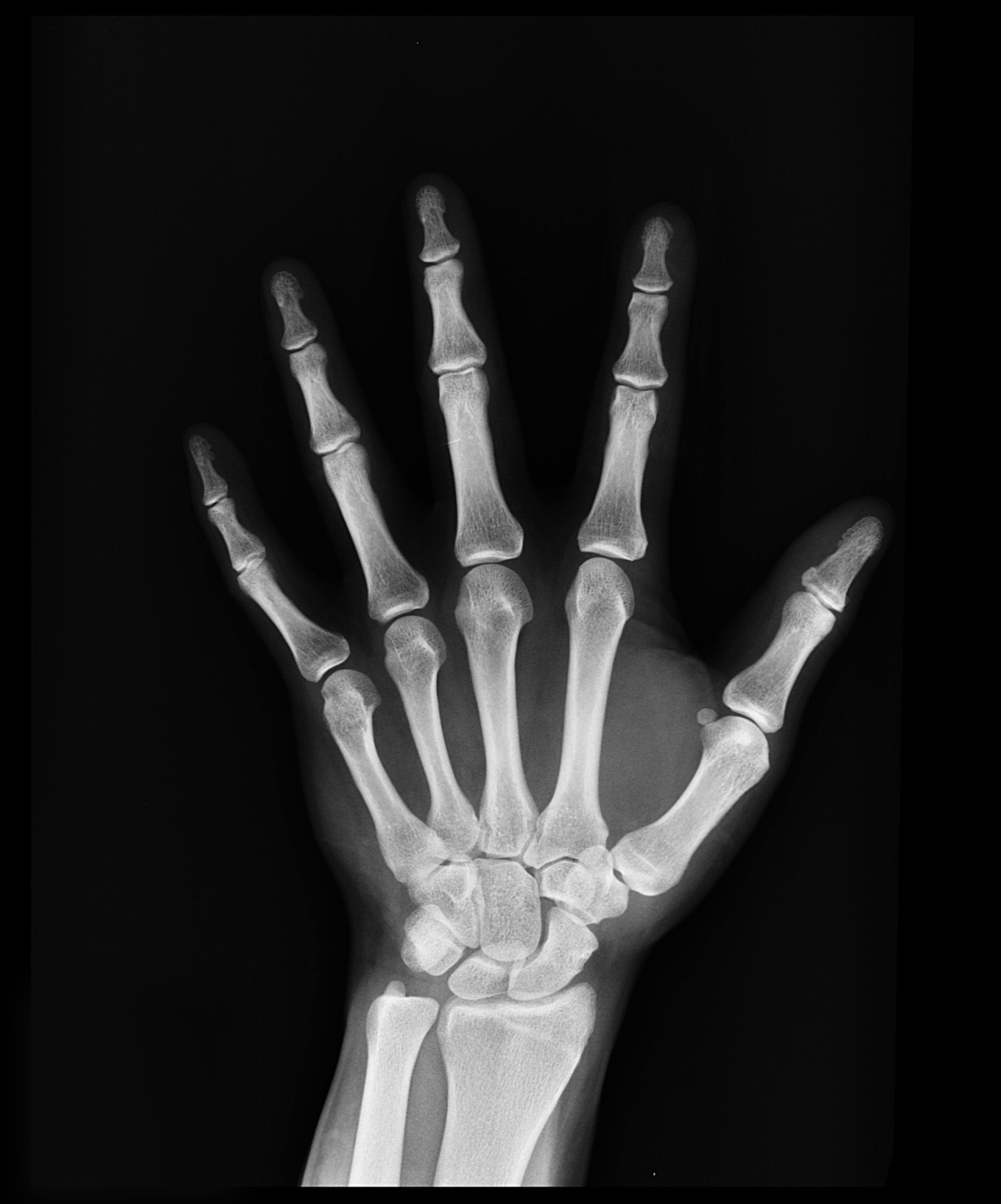
X Ray Picture Of Hand

Hand X Ray

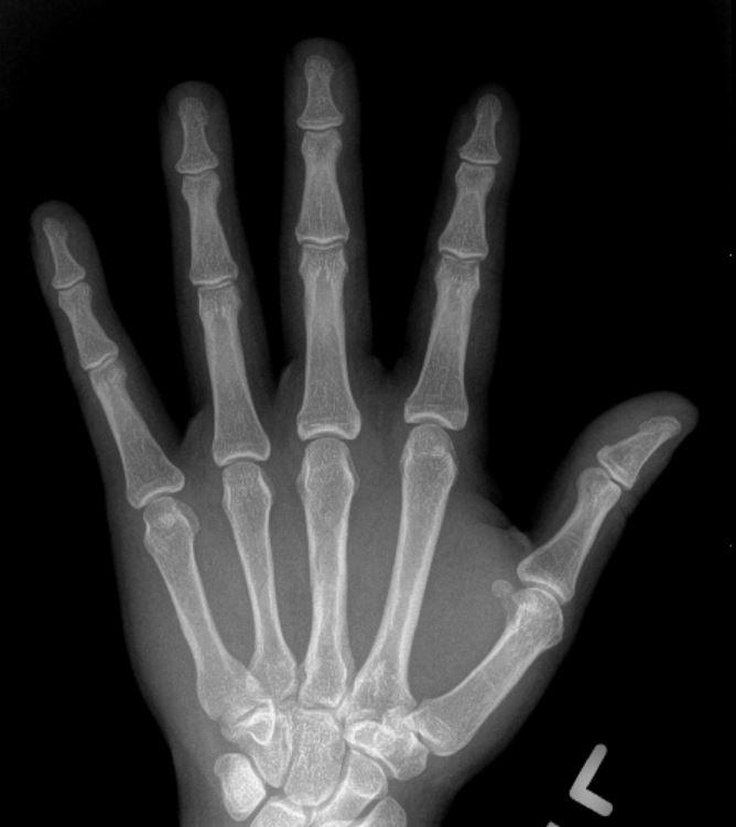
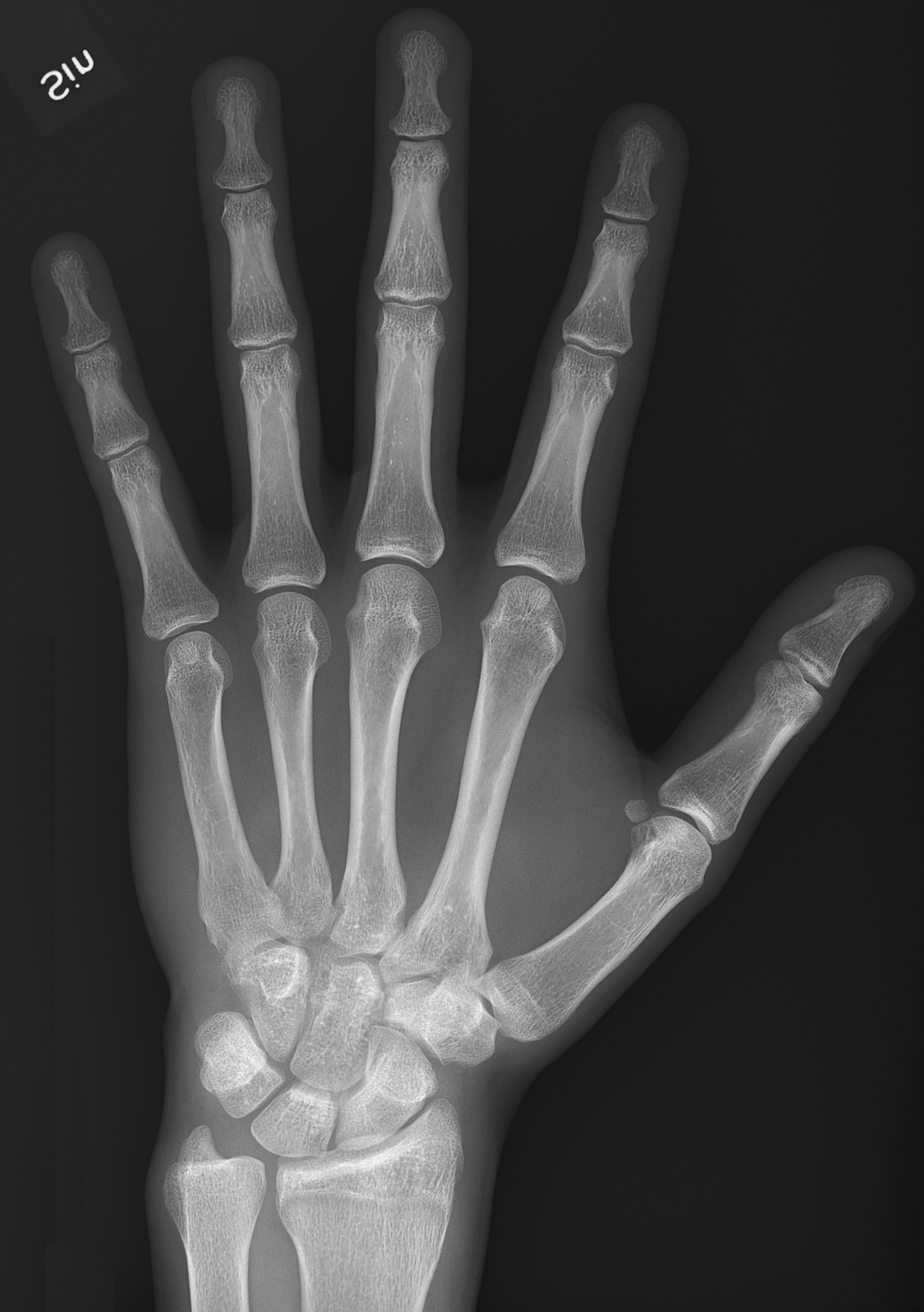
Normal Hand X Ray Of A 19 Year Old Boy D943 144 689

Normal Hand Radiology Case Radiopaedia Org

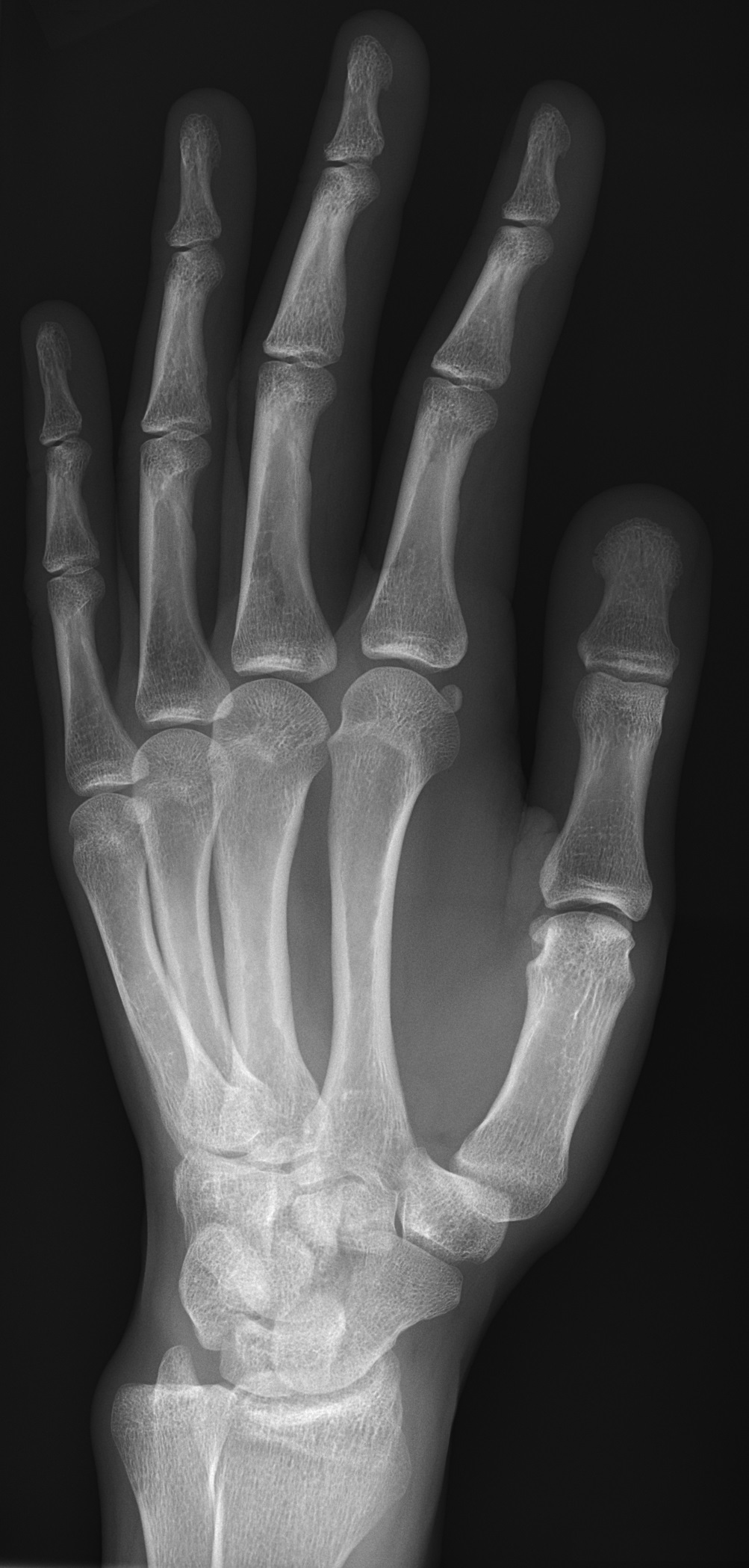
For a pa image the hand lies flat on the x ray plate at the level of the shoulder with the elbow in 90 degrees flexion.
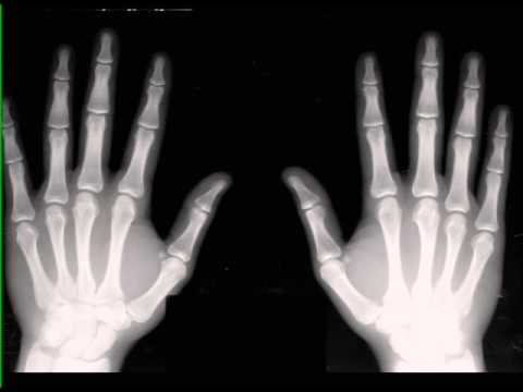
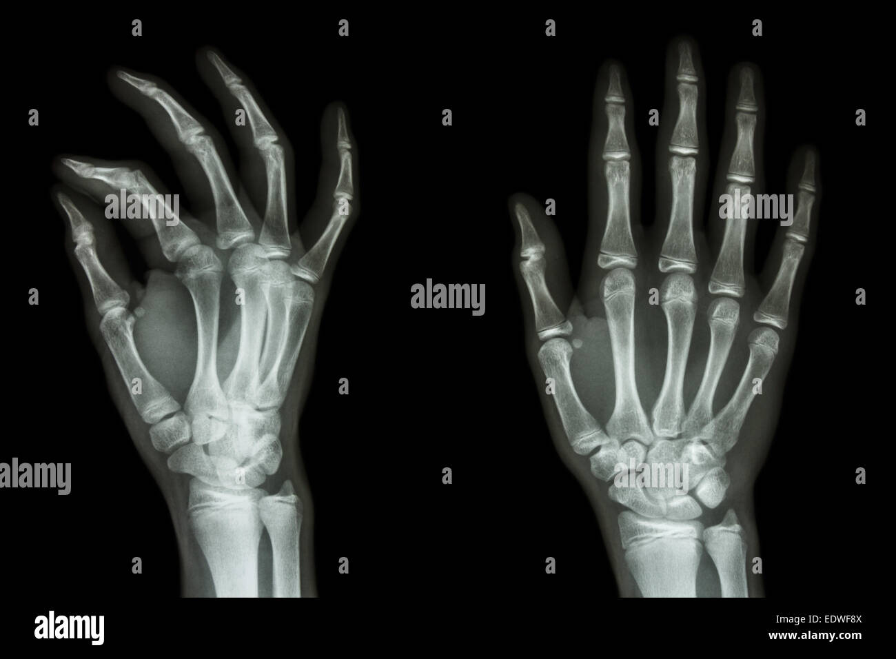
X ray picture of hand. Bones in the handleft and right hand x ray. X ray both hands abnormal of thumb absent of distal and middle phalanges. Narrowing of the space between the bones which are normally covered by cartilage can be a sign of arthritis and its severity. X ray of hand showing all fingers and implantright hand x ray.
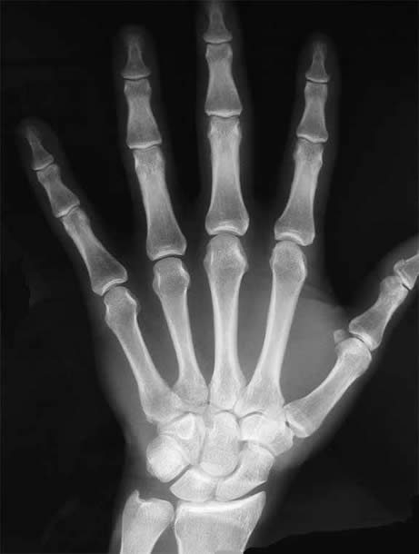
A hand x ray is an imaging technique used to take pictures of hand bones using radiological waves known as x rays for medical purposes. A standard x ray is a simple test in which an x ray beam a form of electromagnetic radiation is passed through the hand to create a two dimensional picture of the bones that form the joint. A doctor can use x rays to view. If you have experienced an acute trauma to your hand finger or wrist and think you may have an injury you should see your doctor.
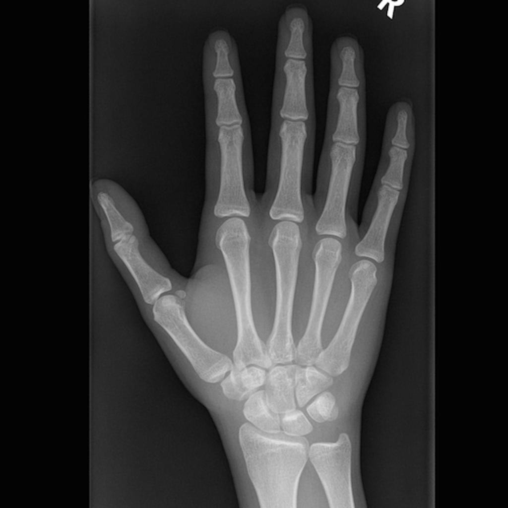
Bones in the handx ray of the right hand. The x ray beam will pass through the hand from dorsal to palmar fig. A left and right hand flat for a diagnostic xrayx ray of right hand with implant. 31797050 film x ray both human hands and arthritis at multiple joint goutrheumatoid.
31797050 film x ray both human hands and arthritis at multiple joint goutrheumatoid. X ray hand on black background. X ray hand many others x ray images in my portfolio. Film x ray right handx ray of the right hand.
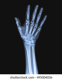
X ray image of both human hands. During the examination an x ray machine sends a beam of radiation through the hand and an image is recorded on special x ray film or a computer. A hand x ray is a safe and painless test that uses a small amount of radiation to take a picture of a persons hand. X ray image of hands making heart symbols.
Hand X Ray Result Free Stock Photo
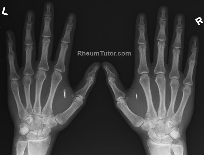
Approach To Hand X Rays Rheumtutor

Approach To Hand X Rays Rheumtutor

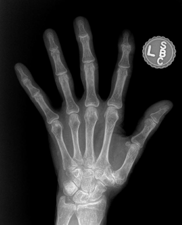
Film X Ray Normal Both Hands Of Child
What Does Hand Arthritis Look Like On X Ray John Erickson Md
X Ray Hand Photos 27 774 X Ray Stock Image Results
File X Ray Of Normal Hand By Dorsoplantar Projection Jpg

Bizarre X Ray Shows Woman Has Pieces Of Gold In Her Hands
Hand Annotated X Rays Radiology Case Radiopaedia Org
What Does Hand Arthritis Look Like On X Ray John Erickson Md

X Hand Startradiology

Bone X Ray
Normal Hands On X Ray
X Ray Normal Hand Stock Photos X Ray Normal Hand Stock

Radiographic Anatomy Hand Ap Medical Anatomy Radiology

Hand X Ray Medical Art Library

X Ray Hand Ap And Oblique View Of The Involved Left Hand

Child S Hand X Ray Stock Image P116 0841 Science
Normal Xray
Hand Bone X Ray

Xray Hand Photos 11 760 Xray Stock Image Results

Baby S Hand X Ray Stock Image P116 0845 Science Photo

X Ray Hand With Ok Symbol

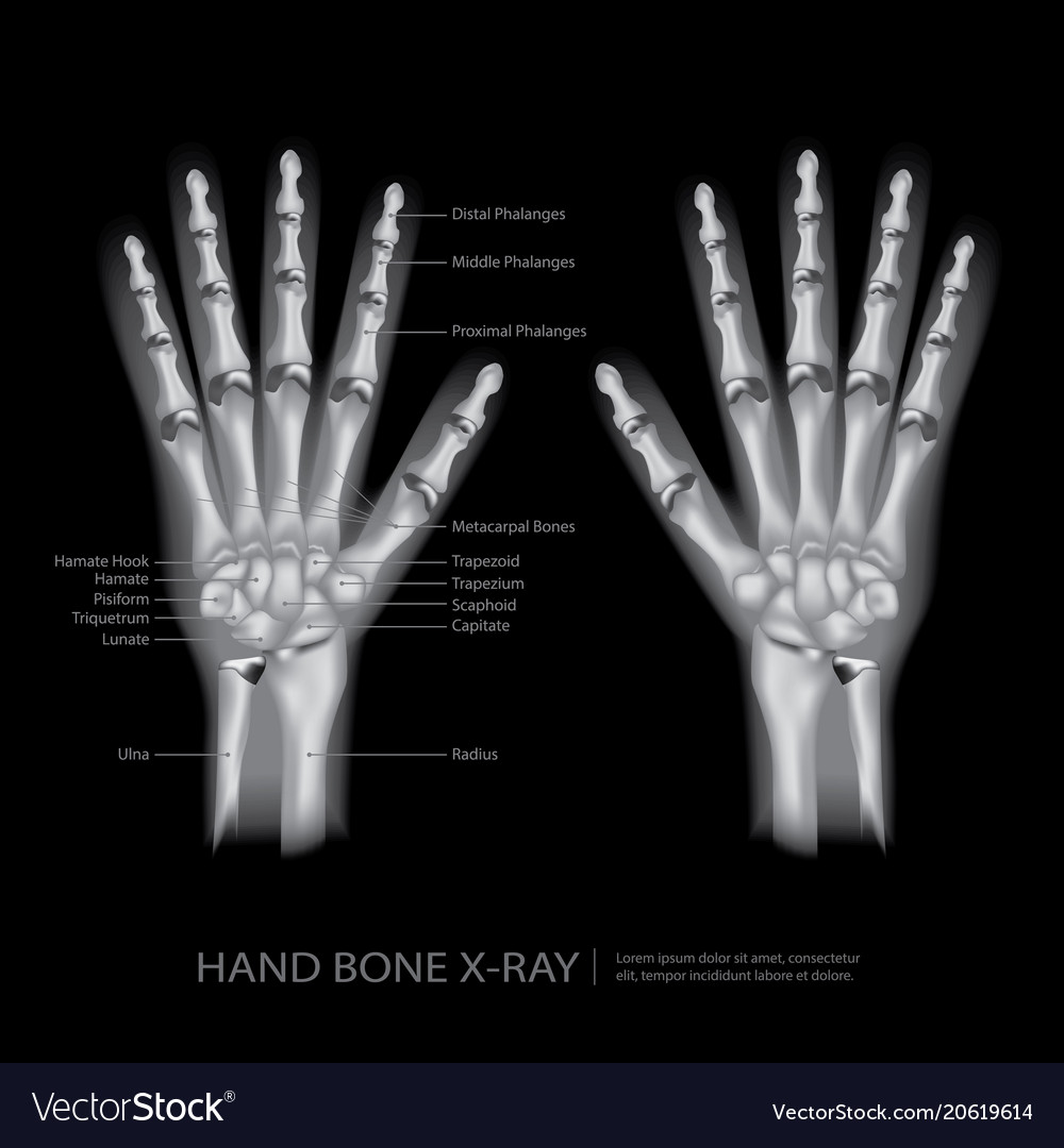
Standard Ap X Ray Of The Hand And Wrist With Labels Naming
File X Ray Of Normal Hand By Oblique Projection Jpg Wikipedia
Adamantium X Ray Hand Bandana

