Microscope Images Of Cells

The Human Body Under The Microscope Discover Magazine
Cellular Organelle Cellular Nucleus Plant Cell

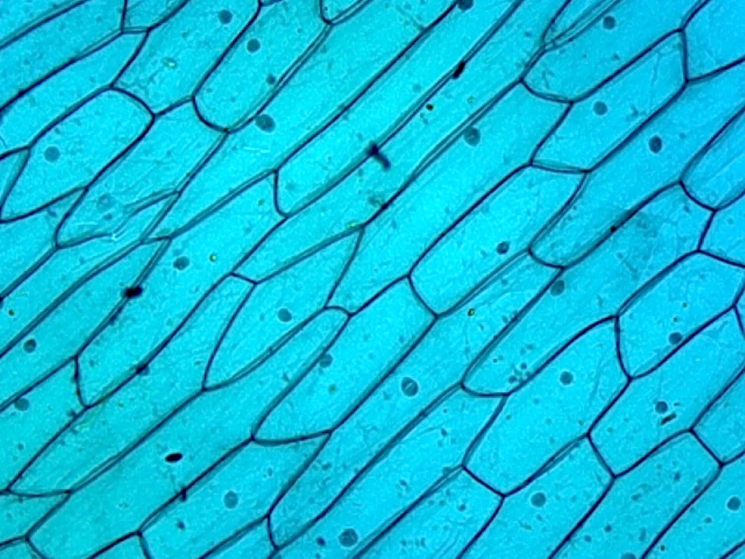
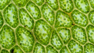
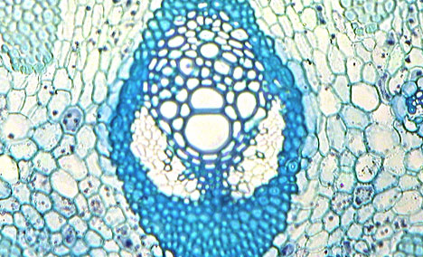
Cell 8 Pictures Of Plant Cells Under A Microscope Plant
Red blood cells erythrocytes are biconcave disc shaped cells that transport oxygen from the lungs to body cells.
Microscope images of cells. Find images of microscope. The cell structure must be adapted and changed according to the function it does in the body. Free for commercial use no attribution required high quality images. A capillary is the smallest type of blood vessel often only just large enough for red blood cells to pass through.
Coloured scanning electron micrograph sem of a red blood cell squeezing out of a torn capillary. With a normal bright field microscope the image is obtained by the simple transmission of light through a cell in culture. This is known as division of labor which is presented in any living organism. Consequently the cell appears as a bright object against a dark background.
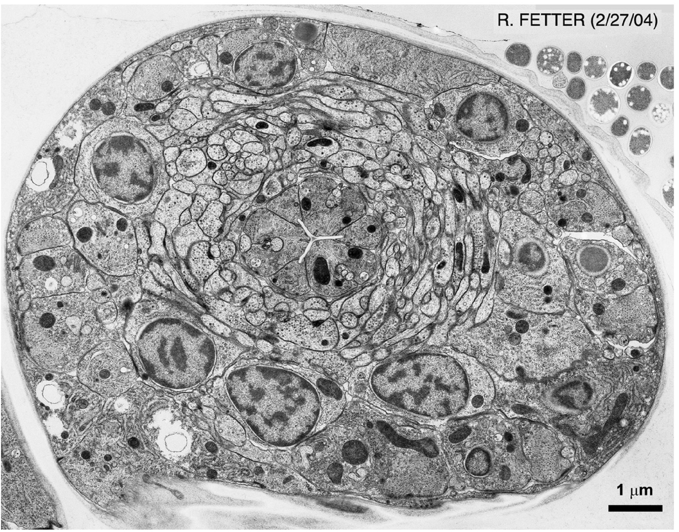
Microscopes provide magnification that allows people to see individual cells and single celled organisms such as bacteria and other microorganisms. Generalized structure of animal cell plant cell under microscope. That is the image was obtained without any fluorophores stains or dyes using only the metabolic co factors of nadh and fad which are already inside of cells. Types of cells that can be viewed under a basic compound microscope include cork cells plant cells and even human cells scraped from the inside of the cheek.
Cell structure hydrilla view of the leaf surface showing plant cells under the microscope for classroom education. This image of a breast cancer tumor and its microenvironment was obtained from a live mouse model using multiphoton microscopy and endogenous fluorescence. Featured image the golgi apparatus. Liposarcoma of a human photomicrograph panorama as seen under the microscope 200x zoom.
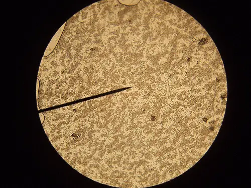
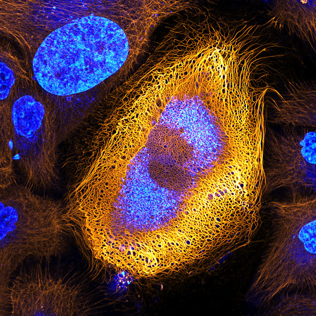
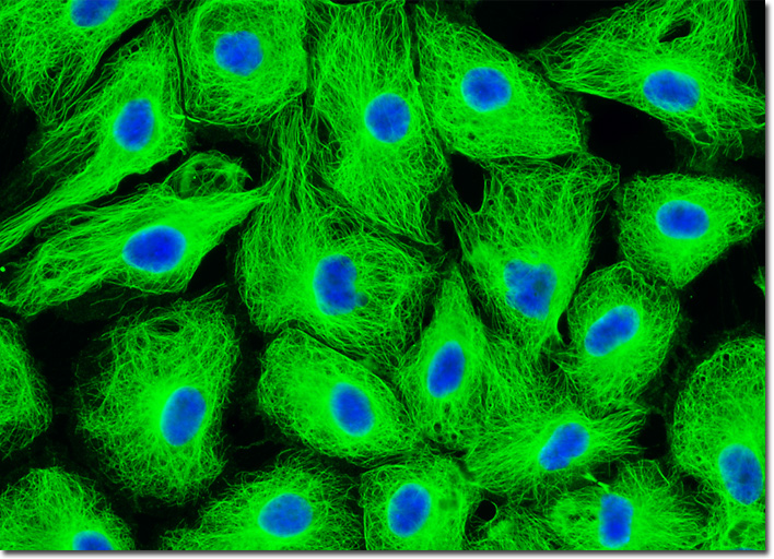
Images of the same cell obtained by four kinds of light microscopy are shown in figure 9 8. Herpes virus or germs microorganism cells under microscope. This is the reason why your palm has a thicker skin unlike the skin of your face. Download cells microscope stock photos.
How To Become A Live Cell Paparazzo A Beginner S Guide

Plant Cells Under Microscope

See Impressive Microscopic Images Of The Human Body
Can We See A Cancer Cell Through A Compound Microscope Quora

Science Plant Cells By Light Microscope
Stunning Microscopic View Of Human Skin Cells Wins 2017

Scanning Electron Microscope Wikipedia
Microscopy Intro To Microscopes How They Work Article

White Blood Cells Crawling Under The Microscope 1000x Magnification

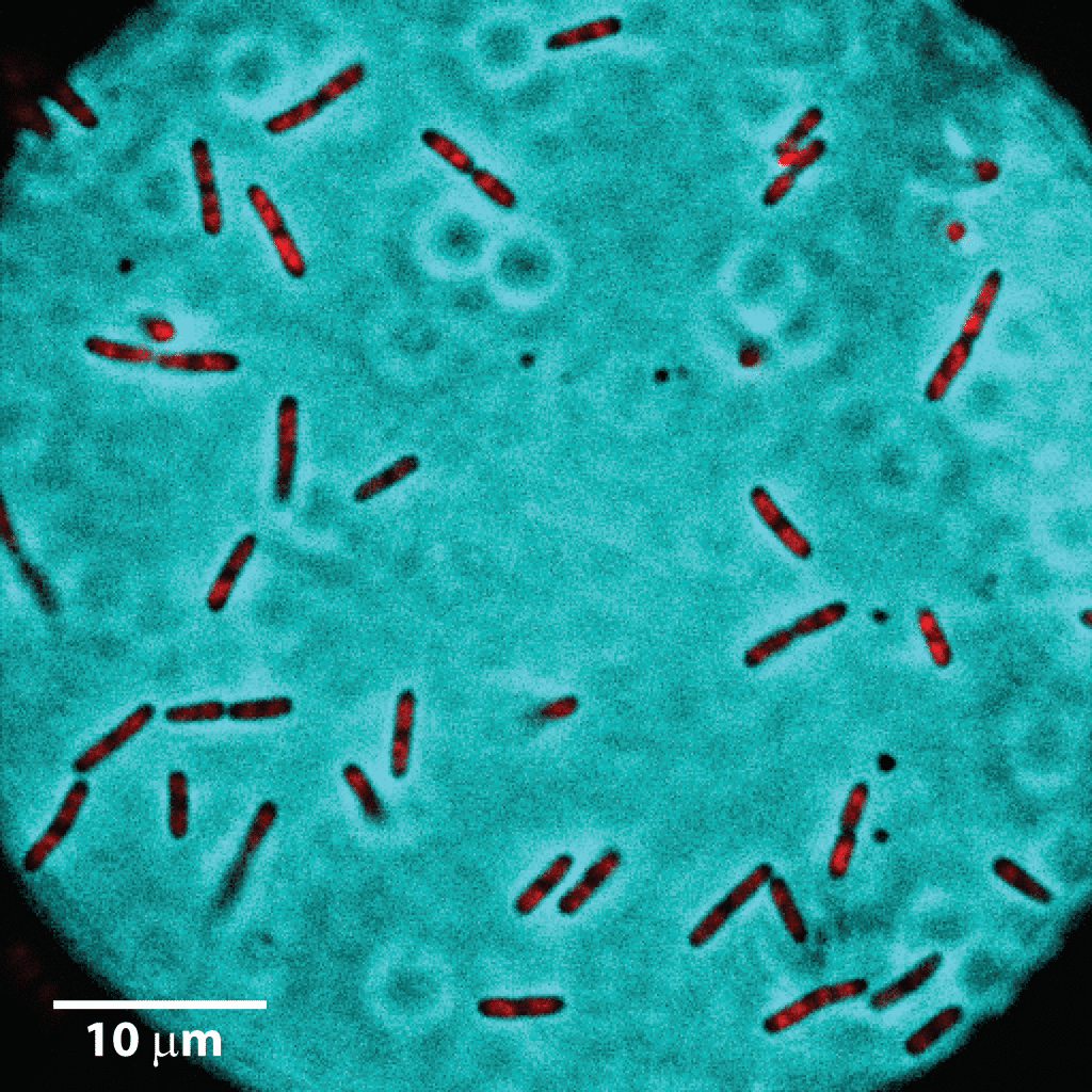
A New Dna Microscope Peers Deep Inside Living Cells Stat

Embryonic Stem Cells Colony Under Stock Footage Video 100 Royalty Free 1023550495 Shutterstock
Some Grass Cells When Viewed Under A Microscope Look Like

Normal Red Blood Cells Under The Microscope

Pin By Ugne Vo On Nature Things Under A Microscope
Cells To Systems Revision 2 Ks3 Biology Bbc Bitesize
Molecular Expressions Microscopy Primer Specialized
Dna Microscopy Offers Entirely New Way To Image Cells
Cell Under Microscope Untamed Science

Electron Microscope Images Show Colourful Cells That Pre

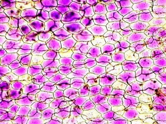
Onion Epidermis With Large Cells Under Microscope
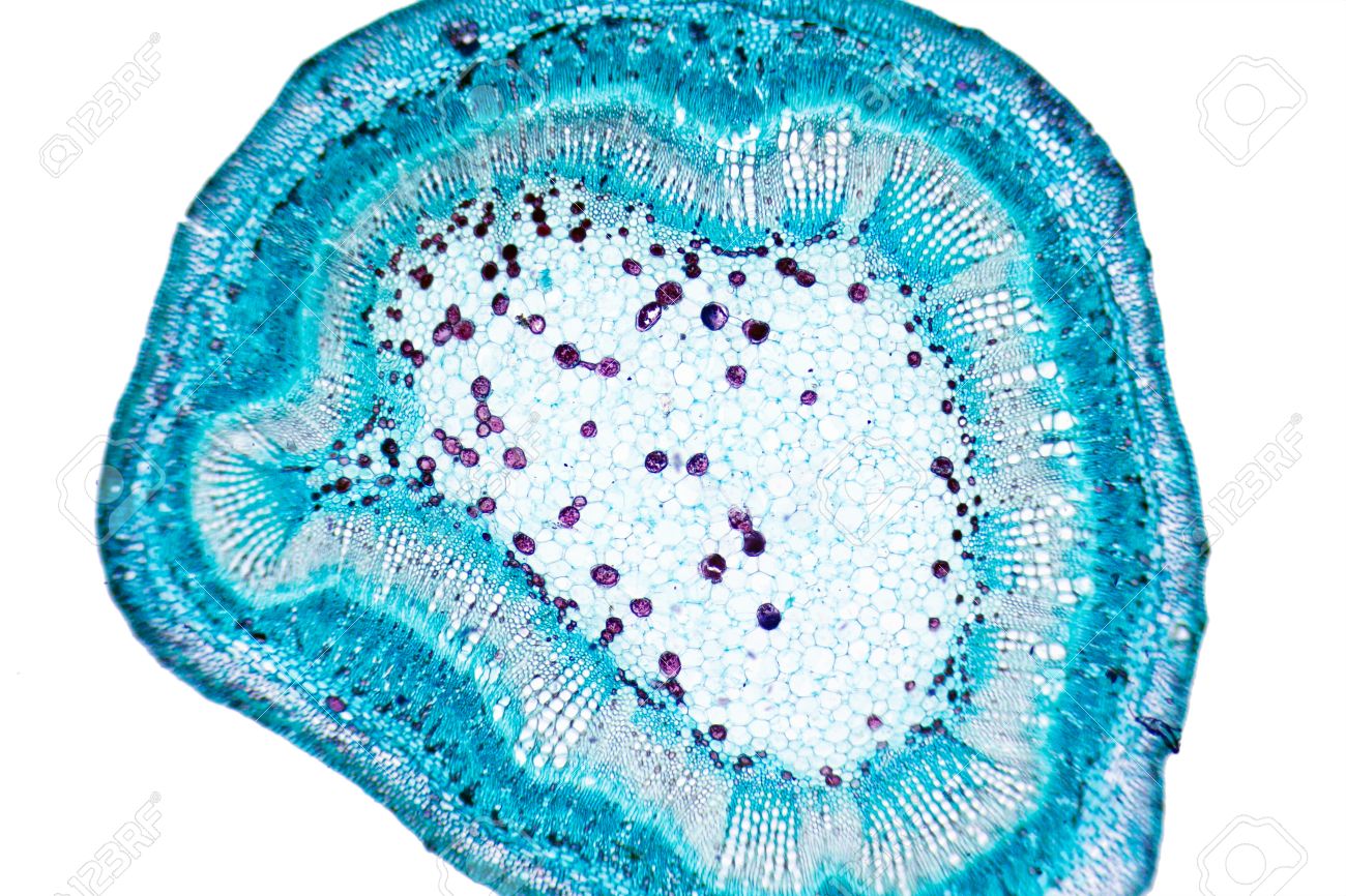
Plant Cells Under Microscope

Even Plant Cells Can Be Art If Seen Under A Microscope

Now In 3d Microscope Offers New Way To View Live Cancer

Microscopic Animal Cells Images Kuhn Photo
Methods In Cell Biology

Life Under The Microscope Of Cells Stock Photo C Ocean Fo