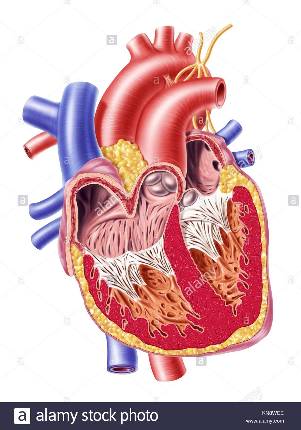
Detailed Picture Of The Human Heart

Human Anatomy Lungs And Heart Heart Anatomy 10 Best Quality

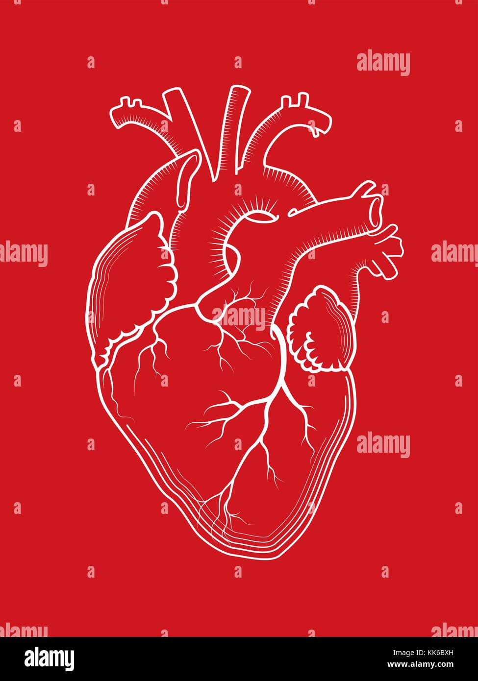
Human Heart Hand Drawn Anatomical Sketch Medicine Vector Color

Xxxl Very Detailed Human Heart Stock Illustration Download

Find high quality human heart stock photos and editorial news pictures from getty images.
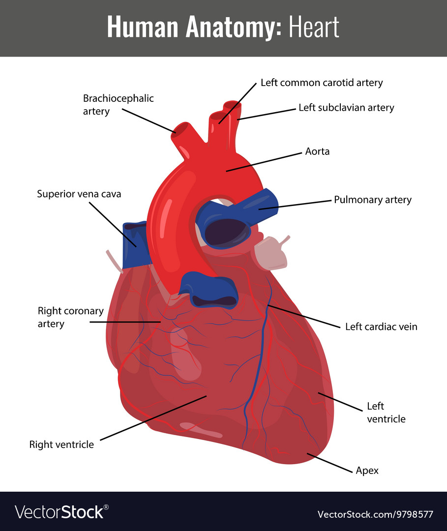
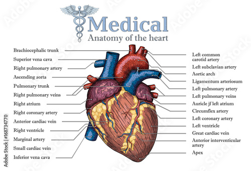
Detailed picture of the human heart. The human heart diagram shows a cross section of a healthy heart and its inside structures. The heart pumps blood through the network of arteries and veins called the cardiovascular system. It is enclosed in a bag like structure called the pericardium and is located between the lungs that is in the middle of the chest behind and slightly to the left of the sternum or breast bone. The human heart resembles the shape of an upside down pear weighing between 7 15 ounces and is little larger than the size of the fist.
The right atrium receives blood from the veins and pumps it to the right ventricle. Oxygenated blood flows into the left ventricle which pumps blood into the arteries that supply the body. The left and right sides of our heart are divided by an internal wall of tissue that is called the septum. Below is a labelled diagram of the human heart with a detailed heart diagram.
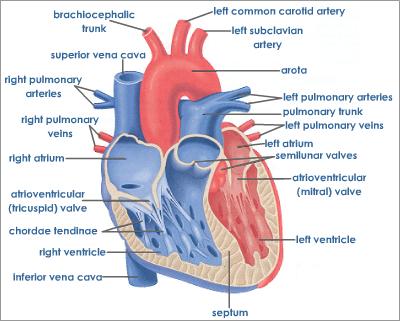
This shows the inside of a normal healthy human heart. Im not going to get into a lot of details about the heart in the post right now because im gonna get more into it later. Veins of the body return blood to the right atrium from there blood flows into the right ventricle which pumps blood to the lungs. The heart has four chambers.
53285304 hand drawn textured romantic poster with human heart and lettering. Download premium images you cant get anywhere else. The conduction system starts with the pacemaker of the hearta small bundle of cells known as the sinoatrial sa node. The adult heart pumps about 1500 to 2000 gallons per day.
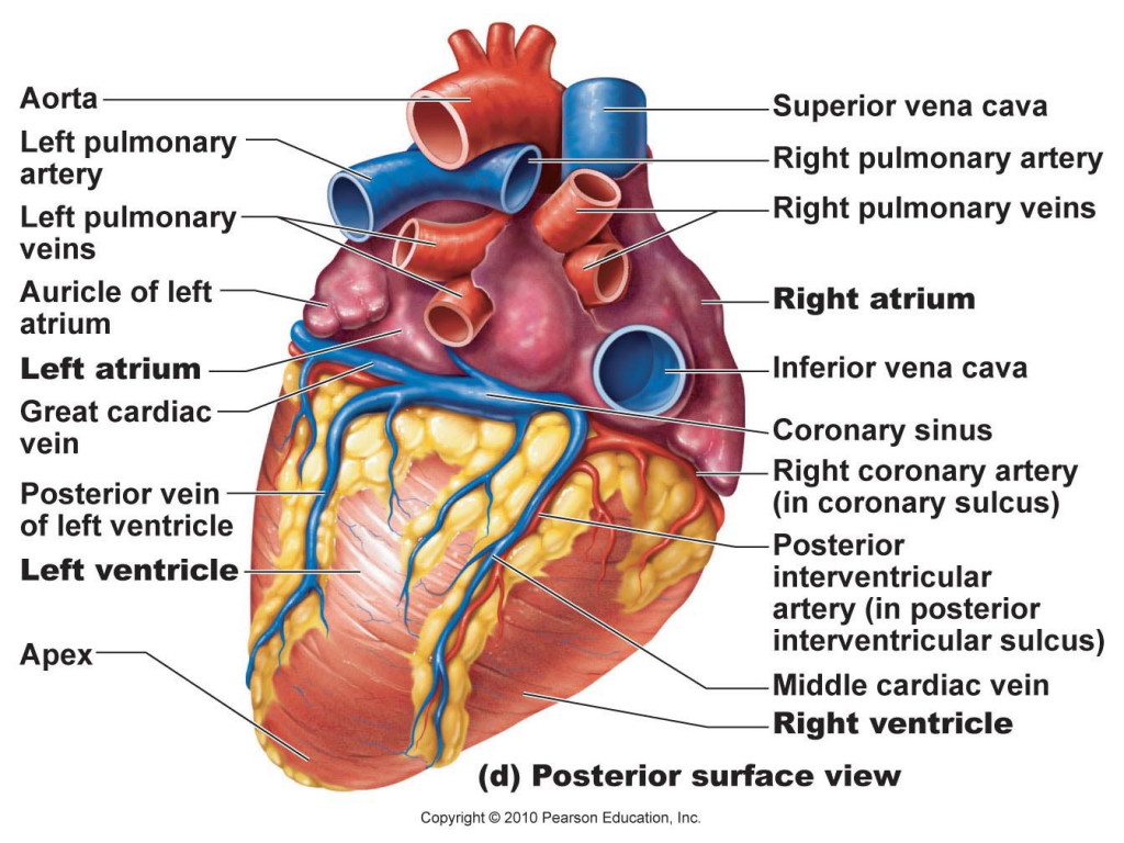
The blue arrow shows the direction in which oxygen depleted blood flows from the body to the lungs. The interior of the heart in human heart diagram. About 1 of the cardiac muscle cells in the heart are responsible for forming the conduction system that sets the pace for the rest of the cardiac muscle cells. The right heart consists of the right atrium which receives deoxygenated blood from the body and the right ventricle which pumps it to the lungs under low pressure.
Below is a picture of the inside of a healthy normal human heart diagram. The heart is a muscular organ about the size of a fist located just behind and slightly left of the breastbone. Chambers of the heart. Picture of heart detail.
Picture of heart detail. After dumping carbon dioxide and picking up oxygen in the lungs the blood goes to the left atrium. And the left heart consisting of the left atrium which receives oxygenated blood from the lung and the left ventricle which pumps it out to the body under high pressure.

Human Heart Detailed Anatomy Isolated On A White Background

Human Heart Anatomy Full Detail Recent Advances In Medical

Human Heart Diagram Detailed Human Heart Diagram Detailed
Human Heart Detailed Anatomy Medical

Realistic Detailed 3d Human Anatomy Heart Healthcare
Free Human Heart Images Download Free Clip Art Free Clip

Realistic Detailed 3d Human Anatomy Heart

Anatomy Of The Human Heart
Human Heart Cross Section With Detailed Internal Structure

Anatomical Human Heart Engraved Stock Vector Colourbox
Anatomical Human Heart Hand Drawn Poster With Inscription Of

Human Heart Detailed Design Stock Illustration Download

Xxxl Very Detailed Human Heart Metal Print
Anatomy System Human Body Anatomy Diagram And Chart Images

Human Heart Outline Sketch Isolated Anatomical Detailed Organ

Realistic Detailed Human Anatomy Heart Closeup View Cardiovascular Organ A Body Medical Health Care Concept Symbol
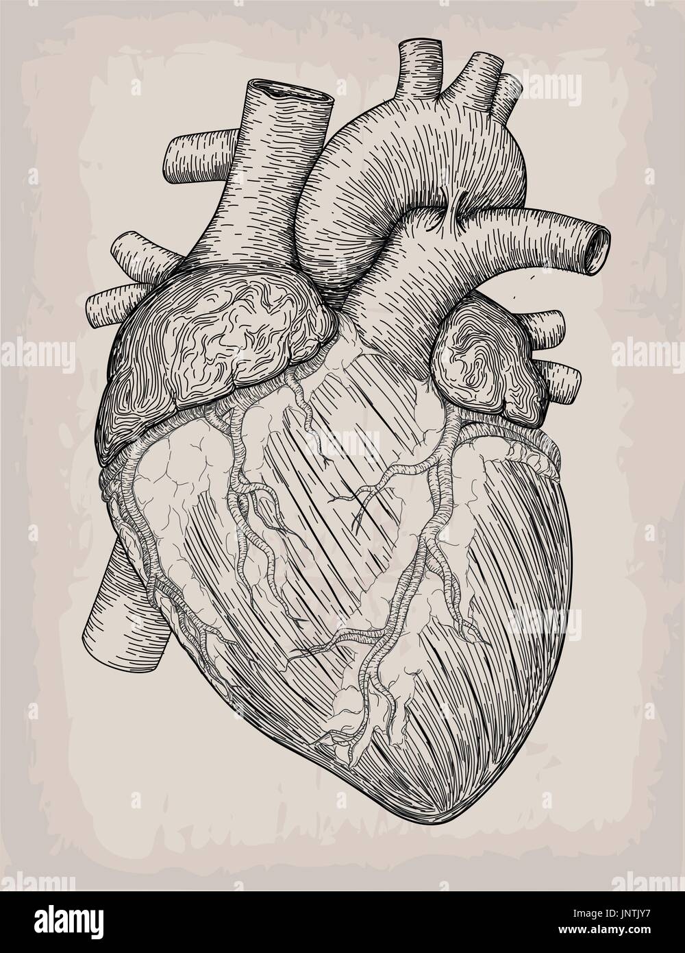
Human Heart Hand Drawn Anatomical Sketch Medicine Vector

Detailed Handdrawn Psychedelic Illustration Human Heart
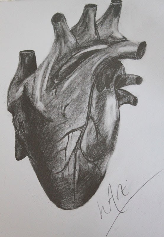
Human Heart Detailed Graphite Drawing Art Time Lapse

Human Heart Drawing Album On Imgur

Human Heart Detailed Design Stock Illustration Download

Anatomical Human Heart Engraved Stock Vector Colourbox
Human Heart Anatomy Icon Detailed Arteries Stock Vector

Axis Scientific Human Heart Model 2 Part Deluxe Life Size Heart Shows 34 Anatomical Internal Structures Held Together With Magnets On Base

Detailed Human Heart Asset Store

Human Heart Detailed